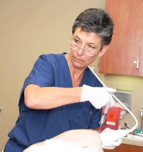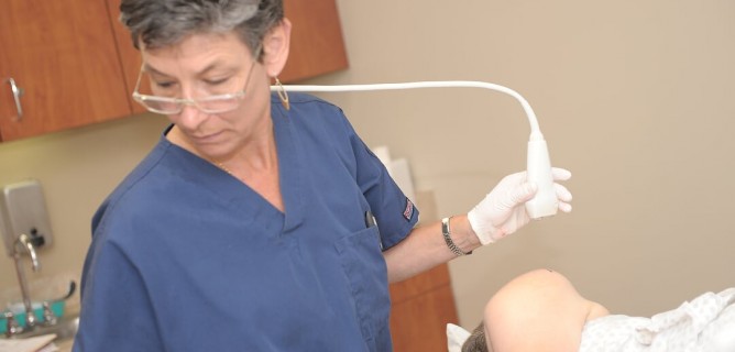
Dr. Annette Zaharoff recently returned from a training on use of ultrasound. She has been utilizing ultrasound for more than 15 years to help patients avoid surgery through effective Regenerative Injection Treatments such as Prolotherapy, PRP and Stem Cell.
By Annette “Dr. Z” Zaharoff, M.D.
I recently returned from a workshop in Orlando, Florida called “Dedicated Ultrasound Training: Hands-on Image Guided Techniques & Applications.”
Ok maybe it doesn’t sound fascinating, but keeping up with how to best use ultrasound technology for the various Regenerative Injection Techniques I employ at the Non-Surgical Center of Texas is critical. I want to remain at the forefront of providing effective Stem Cell, Prolotherapy and PRP Injection Therapy to my patients who could benefit from these treatments.
The training was sponsored by Boston Biolife, a leading provider of accredited hands-on training and education courses for health care providers, with a focus on regenerative medicine. Part of the training was about effectively using ultrasound to locate nerves and determine if there is nerve damage or compression. The workshop also included a possible remedy – ultrasound-guided nerve hydrodissection, a technique using an anesthetic or saline solution to separate the nerve from surrounding fascia or tissue.
Ultrasound has been used in medicine for more than 75 years. When most people think of ultrasound, they often think of the sonogram used to determine the health and sex of a baby during pregnancy. But ultrasound is a very important imaging modality in orthopedics, sometimes referred to as the “orthopedic surgeon’s stethoscope.”
Ultrasound is an amazing diagnostic tool. It can help detect tendon and muscle tears, such as in the shoulder, which can become a problem area from overuse and due to degenerative issues that commonly occur as we age. Ultrasound also can help assess the health of joint and tendon movement and stability. It also makes it possible for physical medicine and rehabilitation specialists like me to get a clearer view of some bone fractures.
Ultrasound equipment also does not emit ionizing radiation like computed tomography (CT) and X-rays, so it’s a totally safe, noninvasive imaging technique. Patients with cardiac pacemakers or metal implants are in no danger from ultrasound scans.
Compared to CT or magnetic resonance imaging (MRI), ultrasound is much more affordable. The portable equipment makes it possible bring the technology into an exam room. I can use ultrasound while my patients are in a comfortable position, without concerns about claustrophobia that may accompany other imaging modalities.
Most importantly for use in Regenerative Injection Treatments, ultrasound provides a very accurate method for administering injections into a joint space or muscle tissue. I can see, in real time, the exact placement of a needle so I can make sure I am addressing the injured area, while ensuring the tip is directed away from neurovascular bundles or other soft tissue.
The technology is also improving. Better transducers, the device that produces the sound waves that help create the sound image (sonogram), are making it possible to be even more accurate with the injections. Studies show that use of ultrasound helps patients to have a better recovery experience in terms of shorter recovery times and improved outcomes.
Ultrasound-guided injections will continue to be the preferred method for regenerative medicine specialists in administering Prolotherapy, Stem Cell and PRP Injections. I’ll continue to keep up with the latest in training to make the most of the tool for the benefit of my patients.
Dr. Annette “Dr. Z” Zaharoff heads the Non-Surgical Center of Texas, focusing on non-surgical treatments to relieve pain and repair injuries. A former professional tennis player who competed on the WTA circuit, Dr. Zaharoff has been utilizing regenerative injection treatments including Stem Cell Therapy, PRP Injection Therapy and Prolotherapy for more than a decade. Learn more about her at www.drzmd.com. You can follow her on Facebook at www.Facebook.com/DrZaharoff.

